Eighteen months ago, artificial intelligence in radiology was mostly conference buzz and vendor demos. Today, if you work CT or MRI at a mid-size to large US facility, you have probably seen at least one AI tool touch your worklist — even if it is still labeled “pilot.” The hype cycle has started to collide with actual shift work, and the gap between the two is worth naming out loud.
This is not a futurist forecast. This is a reality check on what rad tech AI tools are doing right now in November 2026, what they are not doing, and how imaging workflow AI is changing the order in which you scan, the speed at which stat reads happen, and occasionally the questions your radiologists ask before they ever open a study.
If you have been wondering whether the AI radiology 2026 wave is real or just marketing gloss, here is what techs across the country are actually seeing on the floor.
Triage and Worklist Reordering: The Biggest Shift So Far
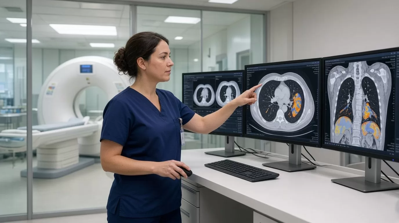
The single most visible change is not a robot doing your job. It is software that reads incoming studies in near-real-time — stroke protocol head CTs, chest CTs for pulmonary embolism, trauma pans — and flags critical findings before the radiologist opens the folder. These triage algorithms typically run in the background the moment reconstruction finishes, sometimes before you have even walked the patient back to the waiting room.
When the algorithm detects a large-vessel occlusion or a saddle PE, it bumps that study to the top of the reading queue and fires an alert to the on-call rad and the ordering physician. At hospitals with stroke or Level I trauma designation, this can cut critical turnaround time by eighteen to twenty-two minutes on average, according to early data shared at the November 2026 RSNA annual meeting in Chicago. That eighteen minutes matters when the interventional team is deciding whether to activate the cath lab at two in the morning.
From a tech perspective, the workflow change is subtle but real. You still perform the scan the same way — same protocol, same positioning, same radiation dose management. But the moment you hit “send,” the AI reads it faster than any human can, and the radiologist may call the ED before you have finished logging the exam. Some techs report feeling a little removed from the loop; others appreciate that critical cases move faster without them having to chase anyone down by phone.
What AI Is Actually Good At (and What It Still Misses)
Most FDA-cleared rad tech AI tools in clinical use today are narrow. They excel at a small number of high-stakes, high-volume tasks:
- Intracranial hemorrhage detection on non-contrast head CT — sensitivity above ninety-five percent in most validation studies, though specificity varies by vendor.
- Pulmonary embolism flagging on chest CTA — reliable for proximal and saddle clots, less consistent with subsegmental filling defects.
- Pneumothorax identification on chest X-ray — helpful in trauma and post-procedure settings, though small apical blebs still get missed.
- Bone age estimation in pediatric radiology — automates a tedious manual task and frees up reading time.
- Lung nodule measurement and tracking on chest CT — consistent diameter calculations across serial studies, reducing inter-observer variation.
What AI still struggles with is context. It can spot a six-millimeter nodule but cannot tell you whether the patient is a lifetime nonsmoker or a two-pack-a-day trucker. It flags every tiny subarachnoid density as possible hemorrhage, generating false positives that radiologists learn to ignore. And it has no intuition about image quality — if your CT is motion-degraded or the contrast bolus mistimed, the algorithm will happily analyze garbage and return confident-looking numbers.
That is why, as of late 2026, every AI finding still requires radiologist over-read. The software assists; it does not replace. The final interpretation, the clinical correlation, and the liability all remain with the physician.
Where Imaging Techs Feel the Impact
For most diagnostic and imaging technologists, day-to-day AI interaction is passive. You are not launching the algorithm manually or adjusting its parameters. It runs automatically when certain exam types hit the PACS, and you see the downstream effects: a re-ordered worklist, a faster callback on a positive study, occasionally a question from the radiologist about whether you repositioned the patient mid-scan because the algorithm flagged asymmetry.
A smaller number of techs — especially those in CT and MRI at academic centers or large health systems — are involved in AI quality assurance workflows. That might mean reviewing flagged cases weekly with the radiology chief to see if the algorithm missed anything, or logging false positives so the vendor can retrain the model. This is new territory, and most imaging schools have not yet added AI literacy to the curriculum. You are learning on the job, often through informal hallway conversations and vendor in-services rather than structured training.
One underappreciated shift: AI-driven worklist prioritization can increase the variability of your scan schedule. If three stroke alerts come in during the same thirty-minute window, you may suddenly have back-to-back stat head CTs even though your original schedule was spaced out. The algorithm does not care about your lunch break. Some departments have started building buffer time into CT and MRI tech schedules to accommodate AI-triggered surges, but that practice is far from universal.
Modality Differences: CT and MRI Lead, Ultrasound Lags
Adoption is uneven across modalities. CT has the most mature AI tools because the images are standardized, cross-sectional, and highly amenable to pattern recognition. MRI is catching up, particularly for brain lesion detection and prostate cancer screening protocols. Mammography has had computer-aided detection for years, though the newest generation uses deep learning and integrates with digital breast tomosynthesis workflows more seamlessly than the old CAD systems.
Ultrasound remains the laggard. The images are operator-dependent, real-time, and hard to standardize, which makes training reliable algorithms much harder. A few AI tools exist for automated ejection-fraction calculation in cardiac echo and for thyroid nodule risk stratification, but they are nowhere near as widespread as CT triage algorithms. If you are an ultrasound tech, AI radiology 2026 probably feels more like 2024 — mostly hype, little hands-on change.
Nuclear medicine and interventional radiology have seen even less AI penetration, though that may shift as dose optimization and real-time guidance tools move out of research labs and into clinical trials.
The Stuff That Is Still Just PowerPoint
Vendor booths at RSNA promise a lot that has not materialized in real hospitals yet. Automated protocoling — where AI selects the CT or MRI sequence based on the clinical indication — is still mostly vaporware. Same with predictive maintenance algorithms that supposedly tell you when your scanner is about to fail. A few sites are piloting these, but they are not standard of care.
Fully automated dose optimization — where the AI adjusts mA and kV in real time based on patient body habitus — exists in high-end scanners, but it is a hardware-integrated feature, not a bolt-on AI product. If your facility bought a CT scanner in 2019, you are probably not seeing that capability unless you upgrade the entire gantry.
And the big one that everyone asks about: Will AI replace imaging techs? The short, evidence-based answer as of late 2026 is no. Positioning patients, selecting the correct protocol, ensuring image quality, managing contrast reactions, and providing compassionate care during a claustrophobic MRI are all tasks that require human judgment, manual dexterity, and emotional intelligence. The AI handles post-acquisition analysis. You handle everything before and during the scan. Those roles are complementary, not competitive.
The bigger workforce risk is not automation; it is understaffing. If hospitals assume AI efficiency gains mean they can run the same imaging volume with fewer techs, that is a management decision, not a technology inevitability. The American Society of Radiologic Technologists has been vocal about protecting staffing ratios even as AI tools proliferate, and that advocacy will matter more than any algorithm.
What This Means for Travel and Per-Diem Imaging Techs
If you work locum or travel contracts in radiology, CT, or MRI, expect facility-to-facility variation in how much AI you encounter. A five-hundred-bed academic medical center in Boston or San Francisco probably has at least two or three FDA-cleared AI tools live in production. A critical-access hospital in rural Wyoming may have heard of AI but is still running a ten-year-old PACS with no budget for add-ons.
That inconsistency creates a small learning curve every time you start a new assignment. Some facilities will give you a thirty-minute AI orientation during onboarding; others will assume you will figure it out by watching the staff techs. Because these tools are still new, there is no standardized training the way there is for, say, GE versus Siemens scanner interfaces. You adapt on the fly.
The good news: AI adoption does not change your core competencies. A strong rad tech who understands anatomy, protocol selection, and patient safety will succeed whether the PACS has an AI layer or not. The algorithm is just another tool in the stack, like automated bolus tracking or 3D reconstruction software. You learn the interface, you trust it when it is right, you catch it when it is wrong, and you move on.
Looking Forward: What to Watch in 2027
Two trends worth tracking as we move into next year. First, interoperability. Right now, most AI tools are siloed — one vendor for stroke detection, another for PE, a third for lung nodules, and none of them talk to each other or integrate cleanly with your EMR. The push toward open standards and HL7 FHIR integration is real, but progress is slow. If that improves, you will see fewer redundant alerts and a cleaner user experience.
Second, reimbursement. Medicare still does not have a clear payment pathway for most AI-assisted radiology reads, which limits how much hospitals are willing to invest. If CMS finalizes new CPT codes or bundled payment models that recognize AI as a billable service, adoption will accelerate. The 2027 Physician Fee Schedule proposed rule is expected in July, and imaging associations are lobbying hard for AI-specific guidance.
For now, the reality on the rad floor is measured and incremental. AI radiology 2026 is not science fiction, but it is not a revolution either. It is a set of narrow, useful tools that make a few high-stakes workflows faster and a little more consistent. The tech job itself — scanning real patients, troubleshooting real equipment, making real-time clinical judgments — remains firmly in human hands.
If you are an imaging tech curious about contracts at facilities with cutting-edge AI workflows or looking for travel assignments where you can build hands-on experience with these tools, reach out to contact@intuites.healthcare with your modality and the regions you are targeting — we can point you toward the hospitals where this technology is live and hiring is active.
Sources
Ready for your next imaging assignment?
Browse open imaging jobs across the US — weekly pay, stipends, and a recruiter who reads every application.