
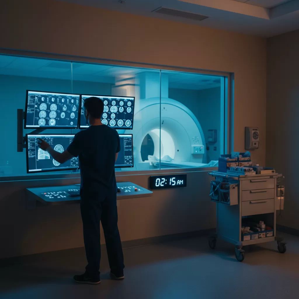
The trauma pager goes off at 2:47 AM. Level One activation. Motorcycle versus guardrail. ETA six minutes.
Your heart rate ticks up — not from panic, but from the controlled adrenaline that comes with high-stakes imaging. You've trained for this. But night shift MRI trauma protocol is different from the daytime version, when you have a full radiology team down the hall and an attending two floors up. Right now, it's you, the ED team, and whatever you pre-staged before the call came in.
This is your complete technical walkthrough for executing flawless MRI trauma workflow when the rest of the hospital is asleep.
Pre-Staging Before the Pager Hits
The best MRI tech trauma responses start hours before the actual activation. During your first hour on night shift — typically that quiet window between 11 PM and midnight — set up your environment for rapid deployment.
Magnet readiness check: Run a quick system QC if your protocol allows it. Verify your gradients are performing within spec, especially if you're on an older 1.5T system that might have drifted during the day. Check your RF coils for any loose connections. You don't want to discover a faulty head coil when a patient with suspected epidural hematoma is already on your table.
Contrast preparation is critical. If your facility uses a power injector for MRI, prime the lines and set your contrast warmer to 98°F. Cold contrast causes more adverse reactions and flows poorly through small-bore IV lines — exactly what trauma patients often have. Keep two pre-filled syringes of gadolinium in your warmer as backup. Some nights you'll get back-to-back traumas, and you won't have time to reload between cases.
Coil staging matters: Position your most common trauma coils where you can grab them in under ten seconds. For most Level One centers, that means:
- 32-channel head coil on the table, centered at iso
- Flex body coils hanging on the left rail
- Spine array underneath the pad, already connected
- Extremity coil on the cart nearest the door
Keep your table at iso-center height with the patient pad already in place. Those 45 seconds you save not adjusting table height and hunting for pads add up when you're trying to get a stroke protocol patient scanned within the therapeutic window.
The Two-Minute Sequence Decision Matrix
When the ED calls with a trauma activation, you need to make sequence decisions before the patient arrives. This is where MRI night shift workflow diverges sharply from CT trauma protocol — you can't just hit “go” on a standard exam.
Ask three questions immediately: mechanism of injury, GCS score, and whether the patient is intubated. Those answers determine your entire scanning strategy.
For head trauma with altered mental status: Your bread-and-butter protocol is sagittal T1, axial T2/FLAIR, axial DWI, axial GRE or SWI, and axial T1 post-contrast if there's any concern for vascular injury. Total scan time: 18-22 minutes on a 3T system. If the patient is combative or GCS is dropping, skip the post-contrast and get the DWI and GRE first — those sequences give you hemorrhage and ischemia, which are the time-sensitive findings.
On a 1.5T magnet, you'll need to adjust. Increase your NEX on DWI from 2 to 4 to maintain adequate signal-to-noise ratio. Your scan time stretches to 28-32 minutes, which means you need to communicate that reality to the ED team upfront. They're used to CT speeds.
Spinal trauma with neuro deficits: Sagittal T1 and T2 of the entire spine, then axial T2 through any area of signal abnormality. If they're worried about ligamentous injury — common in high-speed MVAs — add STIR sequences. These are fat-suppressed and will light up edema in torn ligaments that T2 alone might miss. Position your surface coils before the patient arrives; you won't have time to re-position mid-exam if they're in a cervical collar.
Sequence Modifications for Unstable Patients
Trauma patients decompensate. Your original 22-minute brain protocol might need to become an 8-minute emergent screening exam if their blood pressure drops or they start posturing.
Know your abbreviated protocols cold. Most facilities have a “quick brain” or “emergent stroke” protocol you can adapt: axial DWI, axial GRE, and axial FLAIR. That's it. You get hemorrhage detection and ischemia evaluation in under ten minutes. It's not comprehensive, but it answers the neurosurgeon's immediate question: is there a bleed I need to evacuate right now?
Team Coordination When You're the Only Imaging Tech in the Building
Night shift MRI trauma workflow requires a different communication style than daytime imaging. You don't have a charge tech to run interference or a second MRI tech to help with positioning. You're it.
Establish your role boundaries with the ED and trauma surgery teams during your first week on nights. Make it clear: you will screen every person entering the scan room, you will position the patient, and you will determine if the exam is safe to proceed. Trauma teams are used to moving fast, but magnet safety is non-negotiable.
Pre-scan screening under pressure: When they roll in with a patient, have your screening form ready and make eye contact with the RN. Ask about retained foreign bodies, previous surgeries, and embedded shrapnel — common in penetrating trauma. If the patient is sedated or intubated, physically check for medical devices. Run your handheld metal detector over their chest and abdomen. Ventilators, IV pumps, and monitoring equipment must stay outside the five-gauss line.
Keep your MRI-safe emergency equipment within arm's reach of the magnet. You should have MRI-compatible oxygen, suction, a code cart with non-ferrous tools, and direct communication with the ED. Most night shift MRI traumas don't require code-level intervention, but the ones that do happen fast.
Post-Scan Protocol and Radiologist Communication
You've completed the scan. The patient is stable and heading back to the trauma bay. Your work isn't done.
Upload images immediately and ping the on-call radiologist. Most trauma MRIs on night shift get preliminary reads from teleradiology services, but critical findings — acute epidural hematomas, cord compression, vascular dissections — need a phone call. Don't rely on the PACS notification system alone.
Document your scan parameters and any sequence modifications in your technologist notes. If you abbreviated the protocol due to patient instability, note the reason and the time. If you had to repeat sequences due to motion, document that too. These details matter for quality assurance and for the radiologist's interpretation.
Restock your contrast, clean your coils, and reset your table for the next case. On busy trauma nights, you might get three activations between midnight and 6 AM. The only way to maintain quality is to reset completely between patients.
Maintaining Scan Quality Under Time Pressure
Speed matters in trauma imaging, but not at the expense of diagnostic quality. A motion-degraded DWI sequence is useless for stroke evaluation. A GRE with susceptibility artifact from dental hardware might miss a small hemorrhage.
Use your positioning skills to minimize motion. Strap the head coil snugly — not tight enough to cause pain, but firm enough to prevent small movements. Pad under the knees for spine imaging to reduce lower back discomfort that causes position shifts. If the patient is agitated, consider whether IV sedation is safer than attempting a scan that will need to be repeated.
Monitor your images in real-time. Most modern MRI systems let you preview sequences as they acquire. If you see motion artifact developing, pause and reposition rather than completing a non-diagnostic sequence. Those two minutes you spend fixing the problem save twenty minutes of repeat scanning.
Communication with the patient — even if they're only semi-conscious — makes a difference. Explain what's happening, how long each sequence lasts, and remind them to stay still. Trauma patients are scared and in pain. A calm voice from the intercom helps.
Mastering MRI trauma protocol on night shift takes repetition, but the framework is straightforward: prepare your environment before the emergency arrives, make quick sequence decisions based on clinical information, maintain safety standards under pressure, and never compromise on image quality. When you execute this workflow consistently, you become the MRI tech that trauma surgeons and ED physicians trust — the one they know will deliver diagnostic images when it matters most. If you're looking for night shift MRI positions where advanced trauma imaging is part of the daily workflow, email contact@intuites.healthcare with your experience level and the types of facilities you're targeting.
Ready for your next imaging assignment?
Browse open imaging jobs across the US — weekly pay, stipends, and a recruiter who reads every application.